← sakibchowdhury131.github.io

CovTANet: A Hybrid Tri-Level Attention-Based Network for Lesion Segmentation, Diagnosis, and Severity Prediction of COVID-19 Chest CT Scans

Tanvir Mahmud  ·  Md. Jahin Alam  ·  Sakib Chowdhury  ·  Shams Nafisa Ali  ·  Md. Maisoon Rahman  ·  Shaikh Anowarul Fattah  ·  Mohammad Saquib
Bangladesh University of Engineering and Technology (BUET) & The University of Texas at Dallas
IEEE Transactions on Industrial Informatics, Vol. 17, No. 9 · 2021
Paper (PDF) DOI BibTeX

Abstract

Rapid and precise diagnosis of COVID-19 is one of the major challenges faced by the global community to control the spread of this overgrowing pandemic. In this article, a hybrid neural network is proposed, named CovTANet, to provide an end-to-end clinical diagnostic tool for early diagnosis, lesion segmentation, and severity prediction of COVID-19 utilizing chest computer tomography (CT) scans. A multiphase optimization strategy is introduced for solving the challenges of complicated diagnosis at a very early stage of infection, where an efficient lesion segmentation network is optimized initially, which is later integrated into a joint optimization framework for the diagnosis and severity prediction tasks providing feature enhancement of the infected regions. Moreover, for overcoming the challenges with diffused, blurred, and varying shaped edges of COVID lesions with novel and diverse characteristics, a novel segmentation network is introduced, namely tri-level attention-based segmentation network. This network has significantly reduced semantic gaps in subsequent encoding–decoding stages, with immense parallelization of multiscale features for faster convergence providing considerable performance improvement over traditional networks. Furthermore, a novel tri-level attention mechanism has been introduced, which is repeatedly utilized over the network, combining channel, spatial, and pixel attention schemes for faster and efficient generalization of contextual information embedded in the feature map through feature recalibration and enhancement operations. Outstanding performances have been achieved in all three tasks through extensive experimentation on a large publicly available dataset containing 1110 chest CT-volumes, which signifies the effectiveness of the proposed scheme at the current stage of the pandemic.

Highlights

Method

COVID-19 lesions in chest CT are small, diffused, and irregularly shaped, easily confused with the thorax’s arteries and veins — making both segmentation and, downstream, diagnosis and severity assessment difficult, especially early in infection when lesions are smallest. CovTANet addresses this by first learning to segment lesions precisely, then explicitly feeding those learned lesion features — not just the raw CT volume — into the diagnosis and severity classifiers.
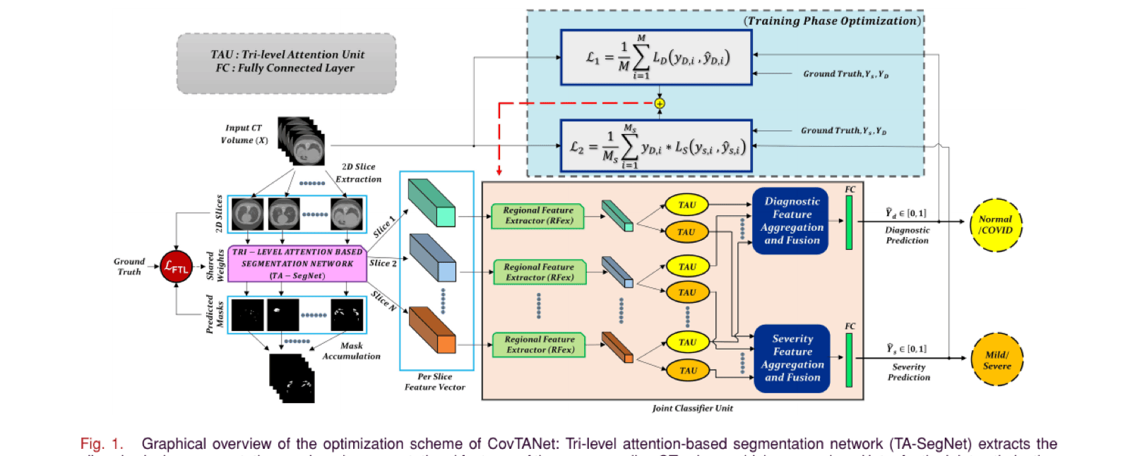
CovTANet overview: TA-SegNet extracts per-slice segmentation masks and feature vectors from the CT volume, which feed a joint classifier unit with tri-level attention units producing diagnostic and severity predictions
Fig. 1 — Overview. TA-SegNet extracts a slicewise lesion mask and a per-slice feature vector from the CT volume. These features pass through a Regional Feature Extractor and multiple Tri-level Attention Units (TAUs) before being aggregated and fused into separate diagnostic and severity predictions.

Tri-Level Attention Unit (TAU)

Each TAU combines three complementary attention mechanisms: channel attention recalibrates which feature channels matter most globally, spatial attention highlights which broad image regions are relevant, and pixel attention further sharpens focus onto the fine-grained pixels most likely to belong to a lesion. Chaining all three lets the network progressively narrow its focus from “which features” to “which regions” to “which exact pixels” matter for the diffused, irregularly-shaped COVID lesions that a single attention mechanism alone tends to miss.

TA-SegNet: Tri-Level Attention Segmentation Network

Built on a U-Net-style encoder–decoder backbone, TA-SegNet places TAUs at both the encoder and decoder stages: encoder TAUs reduce the semantic gap between encoder and decoder feature maps at matching scales, while decoder TAUs refine the reconstructed features with finer spatial detail. A deep fusion scheme combines multiscale features from every encoder–decoder level (rather than relying only on the final decoder output), and an EfficientNet backbone can optionally be used for transfer learning.

Joint Diagnosis & Severity Prediction

Rather than training separate networks per slice, a Regional Feature Extractor (RFEx) processes each slice’s TA-SegNet features, and per-slice TAU-attended features are aggregated across the whole CT volume into two parallel branches — one fused for diagnosis, one for severity — each ending in its own fully-connected classifier. Training happens in two phases: TA-SegNet is optimized first (using a focal Tversky loss suited to the extreme foreground/background imbalance of small lesions), then the full network is jointly optimized for diagnosis and severity using a combined objective — severity loss is only applied to volumes actually diagnosed positive, since severity is undefined for normal cases.
Results

Evaluated on MosMedData (1110 CT volumes from Moscow hospitals; 50 volumes with pixel-level lesion annotations; volumes further labeled normal/mild/severe) with 5-fold cross-validation, plus a secondary 20-volume public segmentation dataset for additional validation.

Segmentation Ablation Study

Starting from a plain U-Net baseline (V1), each proposed component is added incrementally:

VersionEfficientNet BackboneEncoder TAUDecoder TAUEncoder in FusionDecoder in FusionDice (%)
V1 (baseline U-Net)×××××50.5±0.26
V2××××52.4±0.17
V3××××54.9±0.14
V4××××54.6±0.14
V5××××53.4±0.19
V6×××57.1±0.33
V7×60.2±0.26
V8 (TA-SegNet, full)62.3±0.18

Encoder and decoder TAUs each help individually (+4.1% and +2.9% Dice respectively) and more together (+6.6%); fusing multiscale encoder–decoder features adds another +4.4%, combining with two-stage TAUs for +9.7%; and an EfficientNet backbone adds a further +2.1% — each component contributing independently to the final result.

Segmentation vs. Other Networks

Network (MosMedData)Sensitivity (%)Precision (%)Dice (%)IoU (%)
FCN78.8±0.2358.9±0.1635.6±0.3629.3±0.45
U-Net94.3±0.3474.4±0.3250.5±0.2640.3±0.23
VNet84.5±0.4264.6±0.5440.2±0.3336.4±0.26
U-Net++78.1±0.1565.1±0.2537.2±0.2733.3±0.32
CPF-Net82.4±0.2571.3±0.2948.9±0.2137.6±0.38
COPLE-Net85.5±0.1873.1±0.2051.1±0.2141.2±0.38
Mini-SegNet81.5±0.2569.1±0.1943.7±0.2335.2±0.38
Inf-Net92.8±0.2776.9±0.3451.8±0.3141.6±0.27
TA-SegNet (ours)99.6±0.0984.8±0.2662.3±0.1851.7±0.29

TA-SegNet's 99.6% sensitivity means it very rarely misses a lesion outright, while also holding the highest precision of any method tested — most competing networks trade one for the other, producing either excess false positives or missed lesions.

Qualitative Segmentation

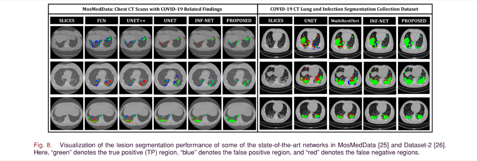
Grid of CT slices with segmentation overlays from FCN, UNet++, UNet, Inf-Net, and the proposed method on MosMedData, and UNet, MultiResUNet, Inf-Net, and the proposed method on a second dataset. Green marks correctly segmented lesion, blue false positives, red false negatives
Fig. 8 — Qualitative segmentation comparison. Green = true positive, blue = false positive, red = false negative. TA-SegNet ("PROPOSED") shows visibly fewer false positives/negatives than FCN, U-Net++, U-Net, Inf-Net, and MultiResUNet across both datasets.

Segmentation on a Secondary Dataset

Network (Dataset-2)Sensitivity (%)Specificity (%)Dice (%)IoU (%)
U-Net75.9±0.3488.9±0.1279.3±0.2674.9±0.18
MultiResUNet77.2±0.3390.3±0.2482.7±0.2877.4±0.15
Attention-UNet81.1±0.2992.2±0.1185.1±0.1479.6±0.25
CPF-Net78.9±0.2791.7±0.1484.4±0.2579.3±0.25
Gated-UNet81.4±0.2492.5±0.1985.6±0.1980.2±0.16
Inf-Net82.7±0.2694.8±0.2186.9±0.3481.1±0.18
TA-SegNet (ours)88.5±0.2298.9±0.1490.2±0.1786.4±0.19

Joint Diagnosis & Severity Prediction

Diagnosis is reported separately for mild and severe cases to highlight early-diagnosis performance, since distinguishing mild COVID from normal CT is the harder and more clinically valuable case:

NetworkDiagnosis: Normal vs. MildDiagnosis: Normal vs. SevereSeverity: Mild vs. Severe
Sen.Spec.Acc.F1Sen.Spec.Acc.F1Sen.Spec.Acc.F1
VGG-1954.463.458.458.663.470.865.966.962.765.561.964.1
ResNet-5061.165.762.563.366.569.369.167.861.163.864.862.4
Xception56.857.959.957.364.867.266.765.962.965.163.163.9
DenseNet12159.764.661.162.165.170.167.867.560.264.460.662.2
InceptionV360.462.159.361.266.669.866.268.262.867.961.465.3
CovTANet (ours)83.890.385.286.993.996.695.894.290.993.491.792.1
All values are percentages (%); ± standard deviation omitted here for space — see Table IV of the paper.

Every baseline here operates directly on raw CT slices; CovTANet's advantage — roughly 20–25 points of accuracy over the best baseline in every column — comes specifically from feeding in TA-SegNet's lesion-focused features instead, letting the classifier ignore the large fraction of each CT volume that is healthy tissue and irrelevant to the diagnosis.

Conclusion

CovTANet shows that segmentation, diagnosis, and severity prediction for COVID-19 chest CT are best addressed together rather than separately: optimizing an attention-based segmentation network first, then explicitly routing its lesion-focused features into the diagnosis and severity classifiers, substantially outperforms networks that classify raw CT volumes directly — particularly on the clinically important case of early, mild-symptom diagnosis. The tri-level (channel, spatial, pixel) attention mechanism proved broadly useful throughout the network, and the resulting framework is proposed as a candidate tool for faster automated mass-screening, with further validation across geographically diverse patient populations suggested as future work.

Citation

@article{mahmud2021covtanet,
  title   = {{CovTANet}: A Hybrid Tri-Level Attention-Based Network
             for Lesion Segmentation, Diagnosis, and Severity
             Prediction of {COVID-19} Chest {CT} Scans},
  author  = {Mahmud, Tanvir and Alam, Md. Jahin and Chowdhury, Sakib
             and Ali, Shams Nafisa and Rahman, Md. Maisoon and
             Fattah, Shaikh Anowarul and Saquib, Mohammad},
  journal = {IEEE Transactions on Industrial Informatics},
  volume  = {17},
  number  = {9},
  pages   = {6489--6498},
  year    = {2021},
  doi     = {10.1109/TII.2020.3048391}
}