The phonocardiogram (PCG) signal is used for the early detection of cardiovascular diseases (CVDs) as it captures the heart sound characteristics. In this article, a spectral attention-based deep learning network is proposed for the automatic detection of cardiac disease from the spectrograms of PCG signals, namely SpectroCardioNet. From a given PCG audio signal, in view of simultaneously utilizing both time- and frequency-domain information, spectrograms, delta-spectrograms, and double-delta-spectrograms are generated. The extracted triple-spectrogram representation is applied in the proposed network as a three-channel 2-D input, where it passes spectral and sequential feature paths. In the spectral feature path, a spectral attention block (SAB) is designed to emphasize some regions in the spectrograms based on a deep attention network and its output is then processed through the spectral pattern detectors (SpPDs). On the other hand, in order to extract the temporal behavior of the frequency components of the spectrograms, a 1-D convolution-based sequential feature extractor is also proposed. Extensive experimentation is carried out on two standard PCG datasets and very satisfactory performance is achieved in comparison to that obtained by some existing methods.
Rather than treat a PCG spectrogram as an ordinary 3-channel color image and reuse a large pretrained vision backbone — the conventional approach — SpectroCardioNet treats it as a single-channel 2-D signal and builds a compact, purpose-built network around it, trained from scratch.
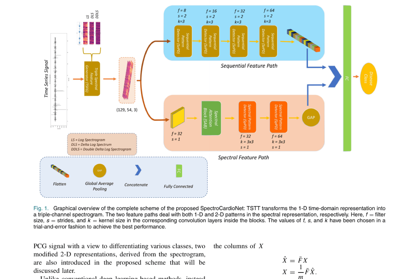
The raw PCG signal is converted to a spectrogram via short-time Fourier transform (256-sample frames, 32ms overlap, at a resampled 2kHz rate — PCG spectral content lives almost entirely below 500Hz). Delta and double-delta spectrograms are then derived to capture how the frequency content changes over time, giving a 3-channel, 2-D input analogous to how delta/delta-delta features are used in speech recognition.
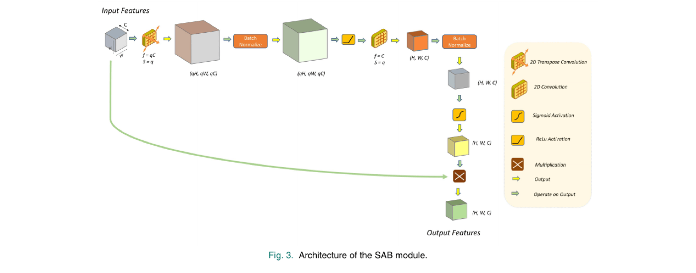
The spectrogram is expanded via transposed convolution, normalized, passed through a convolution that restores the original size, and squashed through a sigmoid into a soft [0,1] mask — upweighting diagnostically relevant time-frequency regions and downweighting the rest. This mask is then multiplied element-wise back onto the original input before further processing.
The SAB’s output passes through two Spectral Pattern Detector (SpPD) blocks — ordinary 2-D convolution, batch normalization, and ReLU — that extract spatial time-frequency correlations, finishing with global average pooling into a 1-D feature vector. In parallel, the sequential feature path runs the un-attended triple-spectrogram through four 1-D Sequential Pattern Detector (SePD) blocks (1-D convolution along the time axis), capturing how frequency-domain features evolve over time. The two resulting feature vectors are concatenated and passed through a fully-connected layer to produce the disease class prediction.
Evaluated on two public PCG datasets: the PhysioNet/CinC 2016 Challenge dataset (3126 recordings, 2 classes — normal/abnormal, tested on the 301 recordings the dataset provider marks as most challenging) and a secondary 5-class dataset (1000 recordings: normal, aortic stenosis, mitral regurgitation, mitral stenosis, mitral valve prolapse).
| Method | Accuracy | Sensitivity | Specificity | F1 |
|---|---|---|---|---|
| SNMFNet | — | 89.18% | 77.66% | — |
| AlexNet+SVM | 85.00% | — | — | — |
| DNN [27] | 85.65% | 86.73% | 85.74% | — |
| DNN [28] | 85.00% | — | — | — |
| TFDDL | 85.16% | — | — | — |
| SpectroCardioNet (ours) | 91.36% | 93.33% | 89.40% | 91.50% |
| Method | Classes | Classifier | Accuracy |
|---|---|---|---|
| WSST | 4 | RF | 95.13% |
| MFCC | 5 | SVM | 91.60% |
| DWT | 5 | SVM | 92.30% |
| IFE | 5 | RF | 94.80% |
| IFE | 5 | KNN | 91.60% |
| SpectroCardioNet (ours) | 5 | DNN | 97.77% |
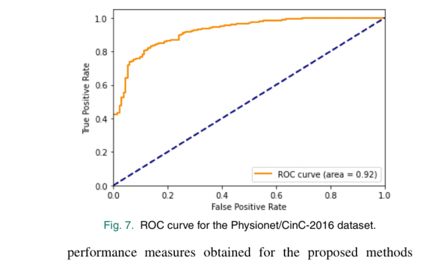
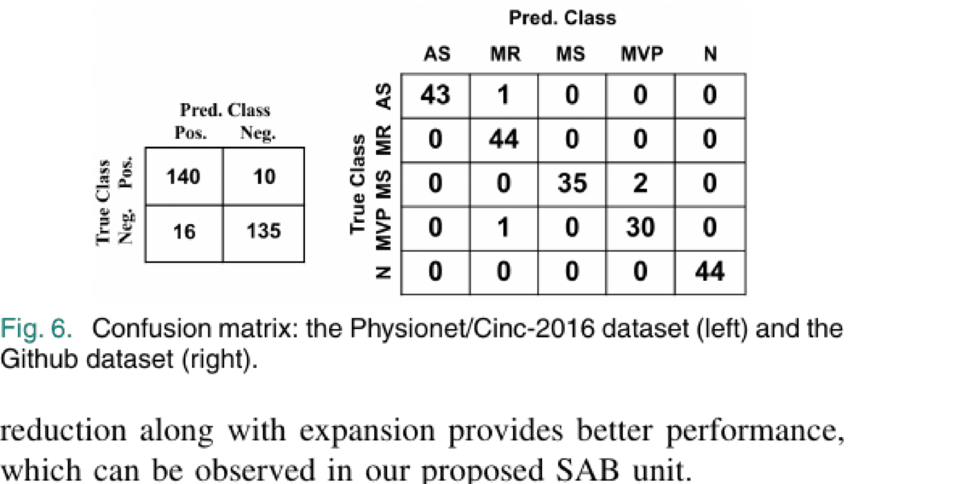
Per-class performance on the 5-class dataset (5-fold cross-validation) averages 97.77% accuracy, 96.57% precision, 96.66% recall, and 96.60% F1 across Normal, Aortic Stenosis, Mitral Regurgitation, Mitral Stenosis, and Mitral Valve Prolapse — with the confusion matrix below showing only a handful of cross-class errors (mostly between mitral stenosis and mitral valve prolapse) and only 5.31% false positives / 3.32% false negatives on the binary PhysioNet task, corresponding to an ROC AUC of 0.92.
SpectroCardioNet shows that a compact, purpose-built network — combining a triple-spectrogram input, a learned spectral attention mask, and parallel spectral/sequential feature paths — can outperform both classical hand-crafted-feature pipelines and larger transfer-learning-based networks on PCG-based heart valve disease detection, while using far fewer parameters and no pretraining. At an estimated 22MB memory footprint, the model is small enough to run on low-power single-board computers, making it a plausible building block for a low-cost clinical diagnostic tool or a remote-monitoring pipeline where a microcontroller streams PCG data to a server running the model.
@article{chowdhury2022spectrocardionet,
title = {{SpectroCardioNet}: An Attention-Based Deep Learning
Network Using Triple-Spectrograms of {PCG} Signal for
Heart Valve Disease Detection},
author = {Chowdhury, Sakib and Morshed, Monjur and Fattah, Shaikh Anowarul},
journal = {IEEE Sensors Journal},
volume = {22},
number = {23},
pages = {22799--22807},
year = {2022},
doi = {10.1109/JSEN.2022.3196263}
}